Skin manifestations of COVID-19 in children: Part 1
D Andina 1, A Belloni-Fortina 2, C Bodemer 3, E Bonifazi 4, A Chiriac 5, I Colmenero 6, A Diociaiuti 7, M El-Hachem 7, L Fertitta 8, D van Gysel 9, A Hernández-Martín 1, T Hubiche 10, C Luca 5, L Martos-Cabrera 1, A Maruani 11, F Mazzotta 4, A D Akkaya 12, M Casals 13, J Ferrando 14, R Grimalt 15, I Grozdev 16, V Kinsler 17, M A Morren 18, M Munisami 19, A Nanda 20, M P Novoa 21, H Ott 22, S Pasmans 23, C Salavastru 24, V Zawar 25, A Torrelo 1; ESPD Group for the Skin Manifestations of COVID-19
Affiliations
Affiliations
- Department of Dermatology, Hospital Infantil Universitario Niño Jesús, Madrid, Spain.
- Pediatric Dermatology Unit, Department of Medicine DIMED, University of Padua, Padua, Italy.
- Department of Dermatology, Hospital Necker Enfants Malades, Paris Centre University, Paris, France.
- Dermatologia Pediatrica Association, Bari, Italy.
- Nicolina Medical Center, Iasi, Romania.
- Department of Pathology, Hospital Infantil Universitario Niño Jesús, Madrid, Spain.
- Dermatology Unit, Bambino Gesù Children's Hospital, IRCCS, Rome, Italy.
- St Parascheva Infectious Diseases Hospital, Iasi, Romania.
- Department of Pediatrics, O. L. Vrouw Hospital, Aalst, Belgium.
- Department of Dermatology, Université Côte d'Azur, Nice, France.
- Department of Dermatology, Unit of Pediatric Dermatology, University of Tours, SPHERE-INSERM1246, CHRU Tours, Tours, France.
- Department of Dermatology, Ulus Liv Hospital, Istanbul, Turkey.
- Department of Dermatology, Hospital Universitari de Sabadell, Barcelona, Spain.
- Department of Dermatology, Hospital Clìnic, Barcelona, Spain.
- Faculty of Medicine and Health Sciences, Universitat Internacional de Catalunya, Barcelona, Spain.
- Department of Dermatology, Children's University Hospital Queen Fabiola, Brussels, Belgium.
- Department of Paediatric Dermatology, Great Ormond Street Hospital for Children, NHS Foundation Trust, London, UK.
- Pediatric Dermatology Unit, Department of Pediatrics and Dermato-Venereology, University Hospital Lausanne and University of Lausanne, Lausanne, Switzerland.
- Department of Dermatology and Sexually Transmitted Diseases, Jawaharlal Institute of Postgraduate Medical Education and Research (JIPMER), Puducherry, India.
- As'ad Al-Hamad Dermatology Center, Kuwait City, Kuwait.
- Department of Dermatology, Hospital San Jose, Bogota, Colombia.
- Division of Pediatric Dermatology, Children's Hospital Auf der Bult, Hannover, Germany.
- Erasmus MC University Medical Center Rotterdam, Sophia Children's Hospital, Rotterdam, The Netherlands.
- Department of Paediatric Dermatology, Colentina Clinical Hospital, Carol Davila University of Medicine and Pharmacy, Bucharest, Romania.
- Department of Dermatology, Dr Vasantrao Pawar Medical College, Nashik, India.
Abstract
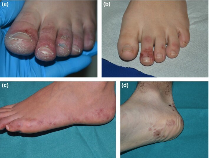
The current COVID-19 pandemic is caused by the SARS-CoV-2 coronavirus. The initial recognized symptoms were respiratory, sometimes culminating in severe respiratory distress requiring ventilation, and causing death in a percentage of those infected. As time has passed, other symptoms have been recognized. The initial reports of cutaneous manifestations were from Italian dermatologists, probably because Italy was the first European country to be heavily affected by the pandemic. The overall clinical presentation, course and outcome of SARS-CoV-2 infection in children differ from those in adults as do the cutaneous manifestations of childhood. In this review, we summarize the current knowledge on the cutaneous manifestations of COVID-19 in children after thorough and critical review of articles published in the literature and from the personal experience of a large panel of paediatric dermatologists in Europe. In Part 1, we discuss one of the first and most widespread cutaneous manifestation of COVID-19, chilblain-like lesions. In Part 2, we review other manifestations, including erythema multiforme, urticaria and Kawasaki disease-like inflammatory multisystemic syndrome, while in Part 3, we discuss the histological findings of COVID-19 manifestations, and the testing and management of infected children, for both COVID-19 and any other pre-existing conditions.
Figures
Similar articles
Skin manifestations of COVID-19 in children: Part 2.
Andina D, Belloni-Fortina A, Bodemer C, Bonifazi E, Chiriac A, Colmenero I, Diociaiuti A, El-Hachem M, Fertitta L, van Gysel D, Hernández-Martín A, Hubiche T, Luca C, Martos-Cabrera L, Maruani A, Mazzotta F, Akkaya AD, Casals M, Ferrando J, Grimalt R, Grozdev I, Kinsler V, Morren MA, Munisami M, Nanda A, Novoa MP, Ott H, Pasmans S, Salavastru C, Zawar V, Torrelo A; ESPD Group for the Skin Manifestations of COVID-19.Clin Exp Dermatol. 2021 Apr;46(3):451-461. doi: 10.1111/ced.14482. Epub 2020 Nov 9.PMID: 33166429 Free PMC article. Review.
Skin manifestations of COVID-19 in children: Part 3.
Andina D, Belloni-Fortina A, Bodemer C, Bonifazi E, Chiriac A, Colmenero I, Diociaiuti A, El-Hachem M, Fertitta L, van Gysel D, Hernández-Martín A, Hubiche T, Luca C, Martos-Cabrera L, Maruani A, Mazzotta F, Akkaya AD, Casals M, Ferrando J, Grimalt R, Grozdev I, Kinsler V, Morren MA, Munisami M, Nanda A, Novoa MP, Ott H, Pasmans S, Salavastru C, Zawar V, Torrelo A; ESPD Group for the Skin Manifestations of COVID-19.Clin Exp Dermatol. 2021 Apr;46(3):462-472. doi: 10.1111/ced.14483. Epub 2020 Nov 18.PMID: 33207021 Free PMC article. Review.
Romita P, Maronese CA, DE Marco A, Balestri R, Belloni Fortina A, Brazzelli V, Colonna C, DI Lernia V, El Hachem M, Fabbrocini G, Foti C, Frasin LA, Guarneri C, Guerriero C, Guida S, Locatelli A, Neri I, Occella C, Offidani A, Oranges T, Pellacani G, Stinco G, Stingeni L, Barbagallo T, Campanati A, Cannavò SP, Caroppo F, Cavalli R, Costantini A, Cucchia R, Diociaiuti A, Filippeschi C, Francomano M, Giancristoforo S, Giuffrida R, Martina E, Monzani NA, Nappa P, Pastorino C, Patrizi A, Peccerillo F, Peris K, Recalcati S, Rizzoli L, Simonetti O, Vastarella M, Virdi A, Marzano AV, Bonamonte D.Ital J Dermatol Venerol. 2023 Apr;158(2):117-123. doi: 10.23736/S2784-8671.23.07539-4.PMID: 37153946
Dondi A, Sperti G, Gori D, Guaraldi F, Montalti M, Parini L, Piraccini BM, Lanari M, Neri I.Eur J Pediatr. 2022 Oct;181(10):3577-3593. doi: 10.1007/s00431-022-04585-7. Epub 2022 Aug 10.PMID: 35948654 Free PMC article. Review.
Colmenero I, Santonja C, Alonso-Riaño M, Noguera-Morel L, Hernández-Martín A, Andina D, Wiesner T, Rodríguez-Peralto JL, Requena L, Torrelo A.Br J Dermatol. 2020 Oct;183(4):729-737. doi: 10.1111/bjd.19327. Epub 2020 Aug 5.PMID: 32562567 Free PMC article.
Cited by
Cutaneous Manifestation in COVID-19: A Lesson Over 2 Years Into the Pandemic.
Danarti R, Limantara NV, Rini DLU, Budiarso A, Febriana SA, Soebono H.Clin Med Res. 2023 Mar;21(1):36-45. doi: 10.3121/cmr.2023.1598.PMID: 37130789 Free PMC article. Review.
SARS-CoV-2 Infection and COVID-19 in Children.
Waghmare A, Hijano DR.Clin Chest Med. 2023 Jun;44(2):359-371. doi: 10.1016/j.ccm.2022.11.014. Epub 2022 Nov 22.PMID: 37085225 Free PMC article. Review.
COVID-19 Associated Vasculitis Confirmed by the Tissues RT-PCR: A Case Series Report.
Belozerov KE, Avrusin IS, Andaryanova LI, Guseva AM, Shogenova ZS, Belanovich IN, Lobacheva AV, Kornishina TL, Isupova EA, Masalova VV, Kalashnikova OV, Nokhrin AV, Panova TF, Dutova YP, Myshkovskaya SL, Kostyunin KY, Komissarov AB, Chasnyk VG, Bregel LV, Kostik MM.Biomedicines. 2023 Mar 13;11(3):870. doi: 10.3390/biomedicines11030870.PMID: 36979849 Free PMC article.
Cazzato G, Ambrogio F, Pisani MC, Colagrande A, Arezzo F, Cascardi E, Dellino M, Macorano E, Trilli I, Parente P, Lettini T, Romita P, Marzullo A, Ingravallo G, Foti C.Vaccines (Basel). 2023 Feb 9;11(2):397. doi: 10.3390/vaccines11020397.PMID: 36851273 Free PMC article. Review.
Ortiz EG, Junkins-Hopkins JM.J Cutan Pathol. 2023 Apr;50(4):321-325. doi: 10.1111/cup.14339. Epub 2022 Oct 18.PMID: 36194075 Free PMC article.
KMEL References
References
-
- Recalcati S. Cutaneous manifestations in COVID‐19: a first perspective. J Eur Acad Dermatol Venereol 2020; 34: e212–13. - PubMed
-
- Mazzotta F, Troccoli T. Acute acro‐ischemia in the child at the time of COVID‐19. Eur J Pediat Dermatol 2020; 30: 71–4.
-
- Cappel JA, Wetter DA. Clinical characteristics, etiologic associations, laboratory findings, treatment, and proposal of diagnostic criteria of pernio (chilblains) in a series of 104 patients at Mayo Clinic, 2000 to 2011. Mayo Clin Proc 2014; 89: 207–15. - PubMed
-
- Ozmen M, Kurtoglu V, Can G et al. The capillaroscopic findings in idiopathic pernio: is it a microvascular disease? Mod Rheumatol 2013; 23: 897–903. - PubMed
-
- Kluger N, Scrivener JN. The use of google trends for acral symptoms during COVID‐19 outbreak in France. J Eur Acad Dermatol Venereol 2020; 34: e358–60. - PubMed
-
- Zhang Y, Cao W, Xiao M et al. Clinical and coagulation characteristics of 7 patients with critical COVID‐2019 pneumonia and acro‐ischemia. Zhonghua Xue Ye Xue Za Zhi 2020; 41: E006. - PubMed
-
- El Hachem M, Diociaiuti A, Concato C et al. A clinical, histopathological and laboratory study of 19 consecutive Italian paediatric patients with chilblain‐like lesions: lights and shadows on the relationship with COVID‐19 infection. J Eur Acad Dermatol Venereol 2020. 10.1111/jdv.16682. - DOI - PMC - PubMed
-
- Mazzotta F, Troccoli T. Erythema multiforme in time of COVID‐19. Eur J Pediatr Dermatol 2020; 2020: 88–9.
-
- He L, Mäe MA, Sun Y et al. Pericyte‐specific vascular expression of SARS‐CoV‐2 receptor ACE2 – implications for microvascular inflammation and hypercoagulopathy in COVID‐19 patients. Med Hypotheses 2020; 144: 110015. - PubMed
