Visual versus semiquantitative analysis of 18F- fluorodeoxyglucose-positron emission tomography brain images in patients with dementia
Affiliations
Affiliations
- Department of Nuclear Medicine, Faculty of Medicine, Kuwait University, Kuwait University, Kuwait.
- Department of Neurology, Faculty of Medicine, Beni-Suef University, Egypt.
- Department of Nuclear Medicine, Ibn Sina Hospital, Kuwait.
- Department of Nuclear Medicine, Adan Hospital, Kuwait.
- Department of Nuclear Medicine, Mubarak Al-Kabeer Hospital, Kuwait City, Kuwait.
- Department of Nuclear Medicine, Faculty of Medicine, Trakya University, Edirne, Turkey.
Abstract
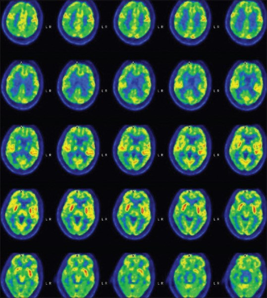
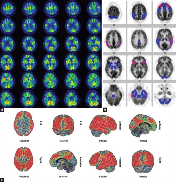
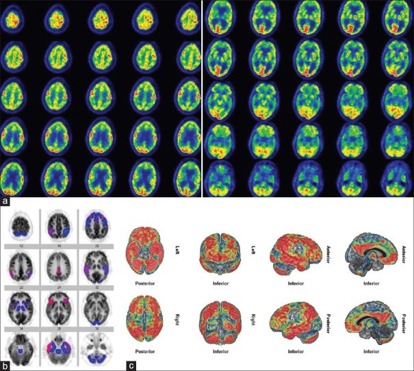
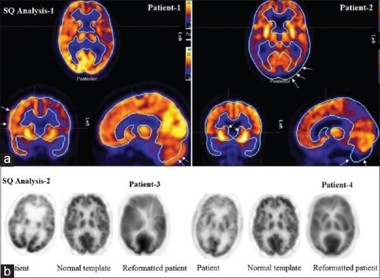
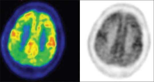
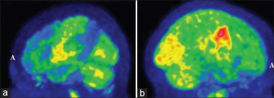
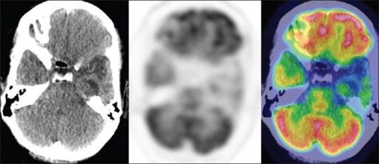
Various studies have reported to the superiority of semiquantitative (SQ) analysis over visual analysis in detecting metabolic changes in the brain. In this study, we aimed to determine the limitations of SQ analysis programs and the current status of 18F- fluorodeoxyglucose (FDG)-positron emission tomography (PET) scan in dementia. 18F- FDG-PET/computed tomography (CT) brain images of 39 patients with a history of dementia were analyzed both visually and semiquantitatively. Using the visually markedly abnormal 18F- FDG-PET images as standard, we wanted to test the accuracy of two commercially available SQ analysis programs. SQ analysis results were classified as matching, partially matching and nonmatching with visually markedly abnormal studies. On visual analysis, 18F- FDG-PET showed marked regional hypometabolism in 19 patients, mild abnormalities in 8 and was normal in 12 patients. SQ analysis-1 results matched with visual analysis in 8 patients (42.1%) and partially matched in 11. SQ analysis-2 findings matched with visual analysis in 11 patients (57.8%) and partially matched in 7 and did not match in 1. Marked regional hypometabolism was either on the left side of the brain or was more significant on the left than the right in 63% of patients. Preservation of metabolism in sensorimotor cortex was seen in various dementia subtypes. Reviewing images in color scale and maximum intensity projection (MIP) image was helpful in demonstrating and displaying regional abnormalities, respectively. SQ analysis provides less accurate results as compared to visual analysis by experts. Due to suboptimal image registration and selection of brain areas, SQ analysis provides inaccurate results, particularly in small areas and areas in close proximity. Image registration and selection of areas with SQ programs should be checked carefully before reporting the results.
Keywords: 18F- fluorodeoxyglucose-positron emission tomography; Brain imaging; dementia; semiquantitative analysis; visual analysis.
Conflict of interest statement
There are no conflicts of interest
Figures
Similar articles
Current Status of 18F-FDG PET Brain Imaging in Patients with Dementia.
Sarikaya I, Sarikaya A, Elgazzar AH.J Nucl Med Technol. 2018 Dec;46(4):362-367. doi: 10.2967/jnmt.118.210237. Epub 2018 Aug 3.PMID: 30076253 Review.
Pianou NK, Stavrou PZ, Vlontzou E, Rondogianni P, Exarhos DN, Datseris IE.Hell J Nucl Med. 2019 Jan-Apr;22(1):6-9. doi: 10.1967/s002449910952. Epub 2019 Mar 7.PMID: 30843003
18F-FDG PET findings in frontotemporal dementia: an SPM analysis of 29 patients.
Jeong Y, Cho SS, Park JM, Kang SJ, Lee JS, Kang E, Na DL, Kim SE.J Nucl Med. 2005 Feb;46(2):233-9.PMID: 15695781 Clinical Trial.
Seki R, Seki H, Toyama T, Koyama K, Endo K, Kurabayashi M.J Cardiol. 2009 Apr;53(2):265-71. doi: 10.1016/j.jjcc.2008.11.009. Epub 2009 Mar 5.PMID: 19304132
Smailagic N, Vacante M, Hyde C, Martin S, Ukoumunne O, Sachpekidis C.Cochrane Database Syst Rev. 2015 Jan 28;1(1):CD010632. doi: 10.1002/14651858.CD010632.pub2.PMID: 25629415 Free PMC article. Review.
Cited by
Franceschi AM, Petrover DR, Giliberto L, Clouston SAP, Gordon ML.World J Nucl Med. 2022 Oct 28;22(1):15-21. doi: 10.1055/s-0042-1757290. eCollection 2023 Mar.PMID: 36923983 Free PMC article.
Isella V, Crivellaro C, Formenti A, Musarra M, Pacella S, Morzenti S, Ferri F, Mapelli C, Gallivanone F, Guerra L, Appollonio I, Ferrarese C.J Neurol. 2022 Aug;269(8):4440-4451. doi: 10.1007/s00415-022-11086-y. Epub 2022 Mar 26.PMID: 35347453 Free PMC article.
KMEL References
References
-
- Matthews FE, Arthur A, Barnes LE, Bond J, Jagger C, Robinson L, et al. A two-decade comparison of prevalence of dementia in individuals aged 65 years and older from three geographical areas of England: Results of the cognitive function and ageing study I and II. Lancet. 2013;382:1405–12. - PMC - PubMed
-
- Prince M, Bryce R, Ferri C. World Alzheimer Report 2011: The Benefits of Early Diagnosis and Intervention. London, UK: Alzheimer's Disease International; 2011.
-
- Rice L, Bisdas S. The diagnostic value of FDG and amyloid PET in Alzheimer's disease-A systematic review. Eur J Radiol. 2017;94:16–24. - PubMed
-
- Donnemiller E, Heilmann J, Wenning GK, Berger W, Decristoforo C, Moncayo R, et al. Brain perfusion scintigraphy with 99mTc-HMPAO or 99mTc-ECD and 123I-beta-CIT single-photon emission tomography in dementia of the Alzheimer-type and diffuse Lewy body disease. Eur J Nucl Med. 1997;24:320–5. - PubMed
-
- Anderson KE, Brickman AM, Flynn J, Scarmeas N, Van Heertum R, Sackeim H, et al. Impairment of nonverbal recognition in Alzheimer disease: A PET O-15 study. Neurology. 2007;69:32–41. - PubMed
-
- Morbelli S, Brugnolo A, Bossert I, Buschiazzo A, Frisoni GB, Galluzzi S, et al. Visual versus semi-quantitative analysis of 18F-FDG-PET in amnestic MCI: An European Alzheimer's disease consortium (EADC) project. J Alzheimers Dis. 2015;44:815–26. - PubMed
-
- Herholz K, Salmon E, Perani D, Baron JC, Holthoff V, Frölich L, et al. Discrimination between Alzheimer dementia and controls by automated analysis of multicenter FDG PET. Neuroimage. 2002;17:302–16. - PubMed
-
- Lehman VT, Carter RE, Claassen DO, Murphy RC, Lowe V, Petersen RC, et al. Visual assessment versus quantitative three-dimensional stereotactic surface projection fluorodeoxyglucose positron emission tomography for detection of mild cognitive impairment and Alzheimer disease. Clin Nucl Med. 2012;37:721–6. - PubMed
-
- Gallivanone F, Della Rosa PA, Castiglioni I. Statistical voxel-based methods and [18F] FDG PET brain imaging: Frontiers for the diagnosis of AD. Curr Alzheimer Res. 2016;13:682–94. - PubMed
-
- Minoshima S, Frey KA, Koeppe RA, Foster NL, Kuhl DE. A diagnostic approach in Alzheimer's disease using three-dimensional stereotactic surface projections of fluorine-18-FDG PET. J Nucl Med. 1995;36:1238–48. - PubMed
-
- Yamane T, Ikari Y, Nishio T, Ishii K, Ishii K, Kato T, et al. Visual-statistical interpretation of (18)F-FDG-PET images for characteristic Alzheimer patterns in a multicenter study: Inter-rater concordance and relationship to automated quantitative evaluation. AJNR Am J Neuroradiol. 2014;35:244–9. - PMC - PubMed
-
- Shivamurthy VK, Tahari AK, Marcus C, Subramaniam RM. Brain FDG PET and the diagnosis of dementia. AJR Am J Roentgenol. 2015;204:W76–85. - PubMed
-
- Herholz K. Guidance for reading FDG PET scans in dementia patients. Q J Nucl Med Mol Imaging. 2014;58:332–43. - PubMed
-
- Bhogal P, Mahoney C, Graeme-Baker S, Roy A, Shah S, Fraioli F, et al. The common dementias: A pictorial review. Eur Radiol. 2013;23:3405–17. - PubMed
-
- Brown RK, Bohnen NI, Wong KK, Minoshima S, Frey KA. Brain PET in suspected dementia: Patterns of altered FDG metabolism. Radiographics. 2014;34:684–701. - PubMed
-
- Lancaster JL, Fox PT, Downs H, Nickerson DS, Hander TA, El Mallah M, et al. Global spatial normalization of human brain using convex hulls. J Nucl Med. 1999;40:942–55. - PubMed
-
- Talairach I, Tournoux P. Co-Planar Stereotaxic Atlas of the Human Brain. New York, NY: Thieme Medical Publishers; 1988.
-
- Galton CJ, Patterson K, Xuereb JH, Hodges JR. Atypical and typical presentations of Alzheimer's disease: A clinical, neuropsychological, neuroimaging and pathological study of 13 cases. Brain. 2000;123(Pt 3):484–98. - PubMed
-
- Brigo F, Turri G, Tinazzi M. 123I-FP-CIT SPECT in the differential diagnosis between dementia with Lewy bodies and other dementias. J Neurol Sci. 2015;359:161–71. - PubMed
-
- Kobylecki C, Langheinrich T, Hinz R, Vardy ER, Brown G, Martino ME, et al. 18F-florbetapir PET in patients with frontotemporal dementia and Alzheimer disease. J Nucl Med. 2015;56:386–91. - PubMed
-
- Sedaghat F, Gotzamani-Psarrakou A, Dedousi E, Arnaoutoglou M, Psarrakos K, Baloyannis I, et al. Evaluation of dopaminergic function in frontotemporal dementia using I-FP-CIT single photon emission computed tomography. Neurodegener Dis. 2007;4:382–5. - PubMed
-
- Engler H, Santillo AF, Wang SX, Lindau M, Savitcheva I, Nordberg A, et al. In vivo amyloid imaging with PET in frontotemporal dementia. Eur J Nucl Med Mol Imaging. 2008;35:100–6. - PubMed
-
- Fodero-Tavoletti MT, Cappai R, McLean CA, Pike KE, Adlard PA, Cowie T, et al. Amyloid imaging in Alzheimer's disease and other dementias. Brain Imaging Behav. 2009;3:246–61. - PubMed
-
- Rowe CC, Ellis KA, Rimajova M, Bourgeat P, Pike KE, Jones G, et al. Amyloid imaging results from the Australian imaging, biomarkers and lifestyle (AIBL) study of aging. Neurobiol Aging. 2010;31:1275–83. - PubMed
-
- Kataoka K, Hashimoto H, Kawabe J, Higashiyama S, Akiyama H, Shimada A, et al. Frontal hypoperfusion in depressed patients with dementia of Alzheimer type demonstrated on 3DSRT. Psychiatry Clin Neurosci. 2010;64:293–8. - PubMed
