Wiener filter improves diagnostic accuracy of CAD SPECT images-comparison to angiography and CT angiography
Affiliations
Affiliations
- Department of Nuclear Medicine and Molecular Imaging, Adan HospitalHadiya, KW.
- Department of Nuclear Medicine, Kuwait Cancer Control Centre, Sabah Medical District, Shuwaikh.
- Department of Nuclear Medicine, Faculty of Medicine, Heath Science Centre, Kuwait University.
- Department of Cardiology, Dabbous Cardiac Centre, Adan Hospital, Hadiya, Kuwait.
Abstract
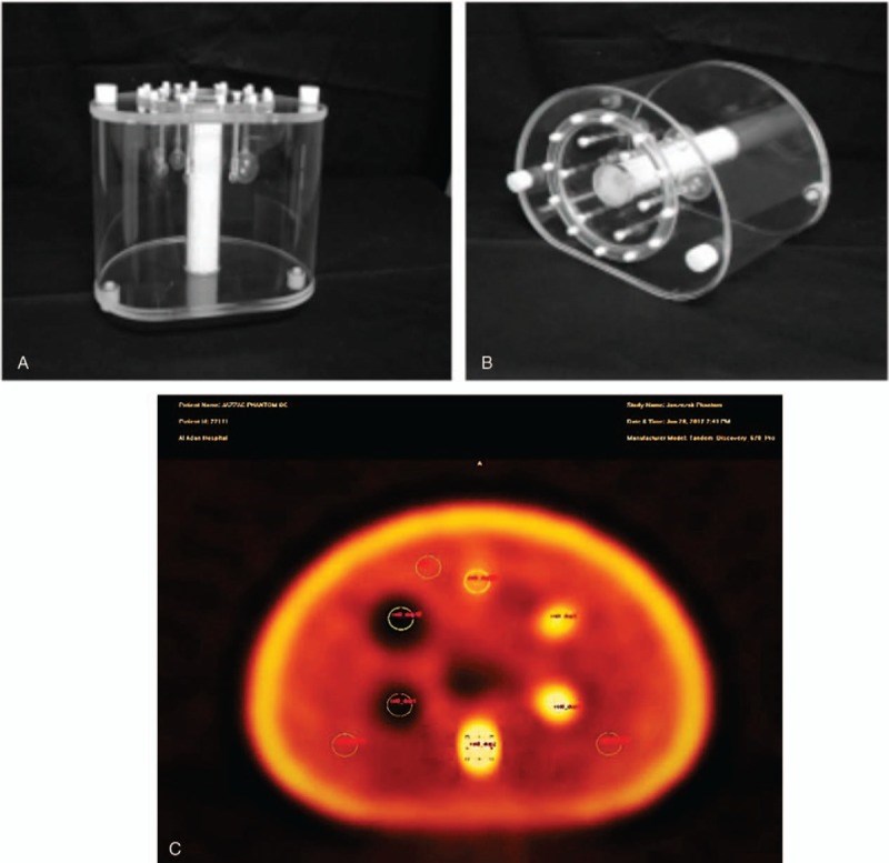
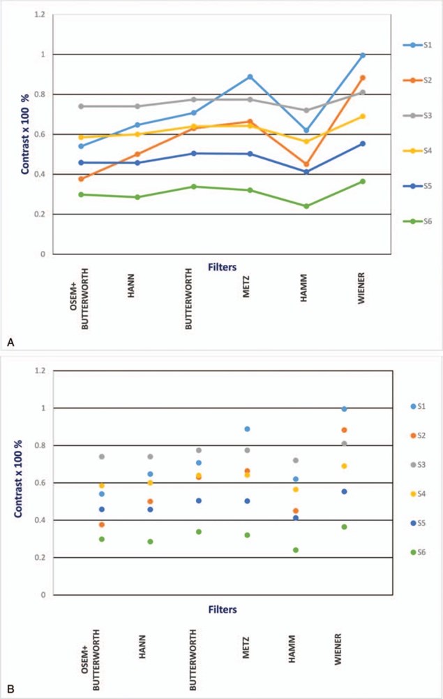
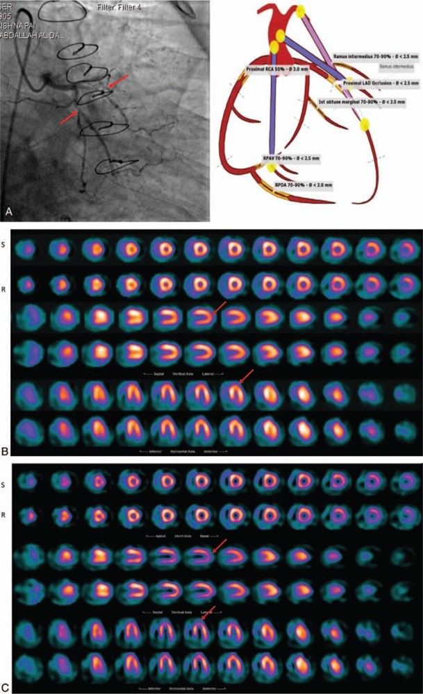
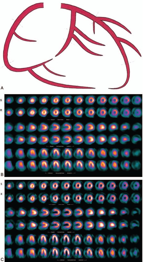
Many discrepancy in selection of proper filter and its parameters for individual cases exists. The authors investigate the impact of the most common filters on patient NM images with coronary artery disease (CAD), and compare the results with the computerized tomography (CT)-Angio and angiography for accuracy.The investigation initiated by performing various single photon emission computerized tomography (SPECT)/CT scan of the national electrical manufacturers association chest phantoms having hot and cold inserts. Data acquired on GE 670 PRO SPECT/CT; 360Ø, 64 frames, 60 seconds, low energy high resolution (LEHR) 128, low energy general purpose (LEGP) with CT attenuation (120 kV and 170 mA). The images reconstructed with filtered back projection and ITERATIVE ordered-subset expectation maximization utilizing filters; Hann, Butterworth, Metz, Hamming, and Wiener. The Image contrast was calculated to assess absolute nearness of the inserts. Based on the preliminary results, then scans of 92 patients with CAD; 64 males and 28 females, age 41 to 77 years old, who had been reported earlier reprocessed with the nominated filter and were reported by 2 NM expert. The results compared to the earlier reports and to the CT-Angio and angiography.The optimization suggested 3 filters; Wiener (Wi), Metz and Butterworth (But) provide the highest contrast (99- 66.4%) and (81- 32%) for the cold and hot inserts respectively, with the (Wi) filter to be the better option. The reprocessed patients scan with the (Wi) presented an elevated diagnostic accuracy, correlated well with the CT-Angio and angiography results (P < .001 and r = 0.79 for [Wi] and P = .004 and r = 0.39 for [But]). The percentage of the false negative for moderate to severe CAD cases reported using Wi filter reduced from 27% to 7% and similarly for mild CAD cases from 7% to 1%.It appears the Wiener filter could produce results with the highest contrast for phantom imaging of various cold and hot spheres and for the patient data which is more consistent with angiography results, with much-elevated accuracy in intermediate cases (r = 0.79 for Wiener and r = 0.39 for Butterworth vs angiography). However, the optimum parameters obtained for the filters have no relation with the resolution of the imaging system, but the details of the objects could be improved.
Conflict of interest statement
The authors have no funding and conflicts of interest to disclose.
Figures
Similar articles
Manrique A, Hitzel A, Gardin I, Dacher JN, Vera P.Nucl Med Commun. 2003 Aug;24(8):907-14. doi: 10.1097/01.mnm.0000084587.29433.ca.PMID: 12869824 Clinical Trial.
Utsunomiya D, Tomiguchi S, Shiraishi S, Yamada K, Honda T, Kawanaka K, Kojima A, Awai K, Yamashita Y.Ann Nucl Med. 2005 Sep;19(6):485-9. doi: 10.1007/BF02985576.PMID: 16248385
Comparison of Low-Pass Filters for SPECT Imaging.
Sayed IS, Ismail SS.Int J Biomed Imaging. 2020 Apr 1;2020:9239753. doi: 10.1155/2020/9239753. eCollection 2020.PMID: 32308670 Free PMC article.
Caobelli F, Akin M, Thackeray JT, Brunkhorst T, Widder J, Berding G, Burchert I, Bauersachs J, Bengel FM.Eur Heart J Cardiovasc Imaging. 2016 Sep;17(9):1036-43. doi: 10.1093/ehjci/jev312. Epub 2015 Nov 30.PMID: 26628617
Performance of 3DOSEM and MAP algorithms for reconstructing low count SPECT acquisitions.
Grootjans W, Meeuwis AP, Slump CH, de Geus-Oei LF, Gotthardt M, Visser EP.Z Med Phys. 2016 Dec;26(4):311-322. doi: 10.1016/j.zemedi.2015.12.004. Epub 2015 Dec 25.PMID: 26725165
Cited by
De Simone L, Chiellino S, Spaziani G, Porcedda G, Calabri GB, Berti S, Favilli S, Stefani L, Santoro G.Children (Basel). 2023 Mar 8;10(3):526. doi: 10.3390/children10030526.PMID: 36980084 Free PMC article.
Rahimian A, Etehadtavakol M, Moslehi M, Ng EYK.Diagnostics (Basel). 2023 Feb 7;13(4):611. doi: 10.3390/diagnostics13040611.PMID: 36832099 Free PMC article.
Rahimian A, Etehadtavakol M, Moslehi M, Ng EYK.J Imaging. 2022 Dec 19;8(12):331. doi: 10.3390/jimaging8120331.PMID: 36547496 Free PMC article.
Kubik A, Budzyńska A, Kacperski K, Maciak M, Kuć M, Piasecki P, Wiliński M, Konior M, Dziuk M, Iller E.PLoS One. 2021 Feb 10;16(2):e0246848. doi: 10.1371/journal.pone.0246848. eCollection 2021.PMID: 33566845 Free PMC article.
KMEL References
References
-
- Hull DM, Peskin CS, Rabinowitz AM, et al. The derivation and verification of a non-stationary, optimal smoothing filter for nuclear medicine image data. Phys Med Biol 1990;35:1641–62. - PubMed
-
- King MA, Long DT, Brill BA. SPECT volume quantitation: influence of spatial resolution, source size and shape, and voxel size. Med Phys 1991;18:1016–23. - PubMed
-
- Fakhri GE, Buvat I, Benali H, et al. Relative impact of scatter, collimator response, attenuation, and finite spatial resolution corrections in cardiac SPECT. J Nucl Med 2000;41:1400–8. - PubMed
-
- Salihin MN, Zakaria A. Determination of the optimum filter for qualitative and quantitative 99mTc myocardial SPECT imaging. Iran J Rad Res 2009;6:173–82.
-
- Global Atlas on Cardiovascular Disease Prevention and Control. Eds: WHO; World Heart Federation; World Stroke Organization 2011, ISBN:979 92 4 1564373.
-
- Shaw LJ, Iskandrian AE. Prognostic value of gated myocardial perfusion SPECT. J Nucl Cardiol 2004;11:171–85. - PubMed
-
- Berman DS, Kang X, Slomka PJ, et al. Underestimation of extent of ischemia by gated SPECT myocardial perfusion imaging in patients with left main coronary artery disease. J Nucl Cardiol 2007;14:521–8. - PubMed
-
- Fujimoto S, Wagatsuma K, Uchida Y, et al. Study of the predictors and lesion characteristics of ischemic heart disease patients with false negative results in stress myocardial perfusion single-photon emission tomography. Circ J 2006;70:297–303. - PubMed
-
- Lue YH, Wacker FJ. Cardiovascular Imaging. London, UK: Manson Publishing; 2010.
-
- Sadremomtaz A, Taherparvar P. The influence of filters on the SPECT image of Carlson phantom. J Biomed Sci Eng 2013;6:291–7.
-
- Hansen CL. Digital image processing for clinicians, part II: filtering. Nucl Cardiol 2002;9:429–37. - PubMed
-
- Boellaard R, Krak NC, Hoekstra OS, et al. Effects of noise, image resolution, and ROI definition on the accuracy of standard uptake values: a simulation study. J Nucl Med 2004;45:1519–27. - PubMed
-
- Salihin MN, Zakaria Z. Relationship between the optimum cut off frequency for Butterworth filter and lung-heart ratio in 99mTc myocardial SPECT. Iran J Radiat Res 2010;8:17–24.
-
- Smith SW. The Scientist and Engineer's Guide to Digital Signal Processing. Santiago, California, USA: California Technical Publishing; 1999.
-
- Gutman F, Gardin I, Delahaye N, et al. Optimisation of the OS-EM algorithm and comparison with FBP for image reconstruction on a dual-head camera: a phantom and a clinical 18F-FDG study. Eur J Nucl Med Mol Imaging 2003;30:1510–9. - PubMed
-
- Knoll P, Mirzaei S, Mullner A, et al. An artificial neural net and error backpropagation to reconstruct single photon emission computerized tomography data. Med Phys 1999;26:244–8. - PubMed
-
- Suzuki S. Spatially limited filters for the two – dimensional convolution method of reconstruction, and their application to SPECT. Phys Med Biol 1992;37:37–52.
-
- Miller TR, Rollins ES. A practical method of image enhancement by interactive digital filtering. J Nucl Med 1985;26:1075–80. - PubMed
-
- Taylor D. Filter choice for reconstruct tomography. Nucl Med Commun 1994;15:857–9. - PubMed
-
- Gilland DR, Tsui BMW, McCartnry WH, et al. Determination of the optimum filter function for SPECT imaging. J Nucl Med 1988;29:643–50. - PubMed
-
- Rajabi H, Pant GS. Optimum filtration for time-activity curves in nuclear medicine. Nucl Med Commun 2000;21:823–8. - PubMed
-
- Sankaran S, Frey EC, Gilland KL, et al. Optimum compensation method and filter cut-off frequency in myocardial SPECT: a human observer study. J Nucl Med 2002;43:432–8. - PubMed
-
- King MA, Gilck SJ, Penney BC, et al. Interactive visual optimization of SPECT pre-reconstruction filtering. J Nucl Med 1987;28:1192–8. - PubMed
-
- Varga J, Bettinardi V, Gilardi MC, et al. Evaluation of pre- and post-reconstruction count dependent Metz filters for brain PET studies. Med Phys 1997;24:1431–40. - PubMed
-
- Tasdemir B, Balci T, Demirel B, et al. Comparison of myocardial perfusion scintigraphy and computed tomography (CT) angiography based on conventional coronary angiography. Nat Sci 2012;4:976–82.
-
- Di Cesare E, Gennarelli A, Di Sibio A, et al. Assessment of dose exposure and image quality in coronary angiography performed by 640-slice CT: a comparison between adaptive iterative and filtered back-projection algorithm by propensity analysis. Radiol Med 2014;119:642–9. - PubMed
-
- Di Cesare E, Gennarelli A, Di Sibio A, et al. Image quality and radiation dose of single heartbeat 640-slice coronary CT angiography: a comparison between patients with chronic atrial fibrillation and subjects in normal sinus rhythm by propensity analysis. Eur J Radiol 2015;84:631–6. - PubMed
-
- Schuijf JD, Bax JJ. CT angiography: an alternative to nuclear perfusion imaging? Heart 2008;94:255–7. - PubMed
-
- Nissen SE. Limitations of computed tomography coronary angiography. J Am Coll Cardiol 2008;52:2145–7. - PubMed
-
- Patel MR. Detecting obstructive coronary disease with CT angiography and noninvasive fractional flow reserve. JAMA 2012;308:1269–70. - PubMed


