Functional Characterization of Posttraumatic Heterotopic Ossification of Tibiofibular Syndesmosis with Dynamic Bone Scan and Single-Photon Emission Computed Tomography/Computed Tomography
Affiliations
Affiliations
- Department of Radiology, Hull University Teachings Hospitals NHS Trust, Hull, HU3 2JZ, United Kingdom.
- Department of Nuclear Medicine, Kuwait Cancer Control Center, Kuwait City, Kuwait.
Abstract
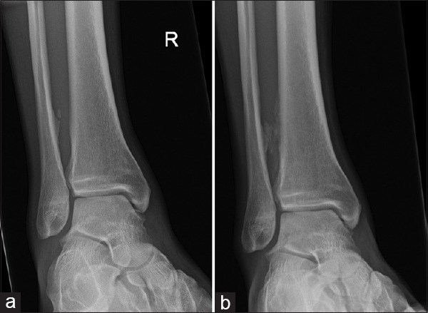
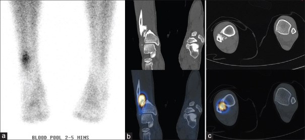
A 53-year-old man was investigated for ongoing right ankle pain and lateral malleolus swelling following a traumatic inversion injury 12 weeks prior. The initial ankle radiograph was normal with no evidence of fracturing. The follow-up radiograph showed bridging ossification in the distal tibiofibular syndesmosis. As the pain did not subside, posttraumatic heterotopic ossification (HO) was suspected, and triple-phase dynamic bone imaging with technetium 99m-methylene diphosphonate was performed to guide further management. The bone scan revealed intense focal tracer activity centered on the HO of the tibiofibular syndesmosis, with no evidence of occult fracturing confirming HO as a pain generator.
Keywords: Dynamic bone imaging; heterotopic bone formation; heterotopic ossification; technetium 99m-methylene diphosphonate; tibiofibular syndesmosis.
Conflict of interest statement
There are no conflicts of interest.
Figures
Similar articles
Botchu R, Douis H, Davies AM, James SL, Puls F, Grimer R.Clin Radiol. 2013 Dec;68(12):e676-9. doi: 10.1016/j.crad.2013.07.020. Epub 2013 Sep 10.PMID: 24034551
Yuan X, Zhang B, Hu J, Lu B.Zhongguo Xiu Fu Chong Jian Wai Ke Za Zhi. 2022 Aug 15;36(8):989-994. doi: 10.7507/1002-1892.202201101.PMID: 35979791 Free PMC article. Chinese.
Veltri DM, Pagnani MJ, O'Brien SJ, Warren RF, Ryan MD, Barnes RP.Foot Ankle Int. 1995 May;16(5):285-90. doi: 10.1177/107110079501600507.PMID: 7633585
Hermans JJ, Beumer A, de Jong TA, Kleinrensink GJ.J Anat. 2010 Dec;217(6):633-45. doi: 10.1111/j.1469-7580.2010.01302.x.PMID: 21108526 Free PMC article. Review.
[Progress in diagnosis and treatment of distal tibiofibular syndesmosis injury].
Liu X, Yu G.Zhongguo Xiu Fu Chong Jian Wai Ke Za Zhi. 2012 May;26(5):612-6.PMID: 22702060 Review. Chinese.
KMEL References
References
-
- !Fu JH, Hwang CC, Chao TH. Tibiofibular synostosis in a military soldier. J Med Sci (Faisalabad, Pakistan) 2003;23:135–8.
-
- Veltri DM, Pagnani MJ, O'Brien SJ, Warren RF, Ryan MD, Barnes RP. Symptomatic ossification of the tibiofibular syndesmosis in professional football players: A sequela of the syndesmotic ankle sprain. Foot Ankle Int. 1995;16:285–90. - PubMed
-
- Tyler P, Saifuddin A. The imaging of myositis ossificans. Semin Musculoskelet Radiol. 2010;14:201–16. - PubMed
-
- Hendifar AE, Johnson D, Arkfeld DG. Myositis ossificans: A case report. Arthritis Rheum. 2005;53:793–5. - PubMed
-
- Botchu R, Douis H, Davies AM, James SL, Puls F, Grimer R. Post-traumatic heterotopic ossification of distal tibiofibular syndesmosis mimicking a surface osteosarcoma. Clin Radiol. 2013;68:e676–9. - PubMed
-
- Kransdorf MJ, Meis JM, Jelinek JS. Myositis ossificans: MR appearance with radiologic-pathologic correlation. AJR Am J Roentgenol. 1991;157:1243–8. - PubMed
-
- Love C, Din AS, Tomas MB, Kalapparambath TP, Palestro CJ. Radionuclide bone imaging: An illustrative review. Radiographics. 2003;23:341–58. - PubMed
-
- Shehab D, Elgazzar AH, Collier BD. Heterotopic ossification. J Nucl Med. 2002;43:346–53. - PubMed
-
- Orzel JA, Rudd TG. Heterotopic bone formation: Clinical, laboratory, and imaging correlation. J Nucl Med. 1985;26:125–32. - PubMed
-
- Drane WE. Myositis ossificans and the three-phase bone scan. AJR Am J Roentgenol. 1984;142:179–80. - PubMed
-
- Vanden Bossche L, Vanderstraeten G. Heterotopic ossification: A review. J Rehabil Med. 2005;37:129–36. - PubMed

