Point-of-Care Ultrasound to Detect Dilated Coronary Sinus in Adults
Affiliations
Affiliations
- Consultant critical care medicine, Internal medicine department, Ahmadi hospital Kuwait.
- 2Critical care Unit, Ahmadi hospital, Kuwait oil company Kuwait.
- 3Assistant Professor, faculty of medicine, Kuwait university Kuwait.
- 4Team leader internal medicine and cardiologist, Ahmadi hospital, Kuwait oil company Kuwait.
Abstract
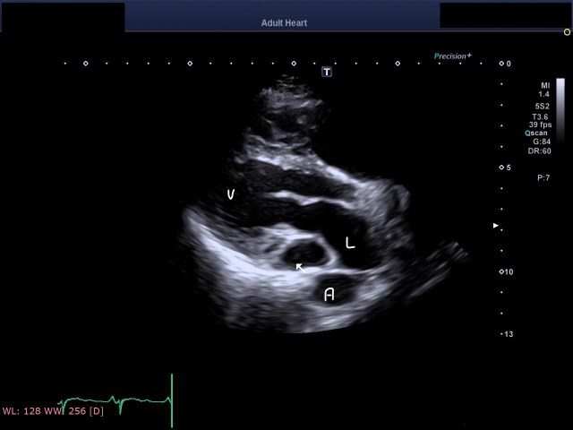
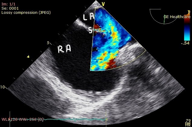
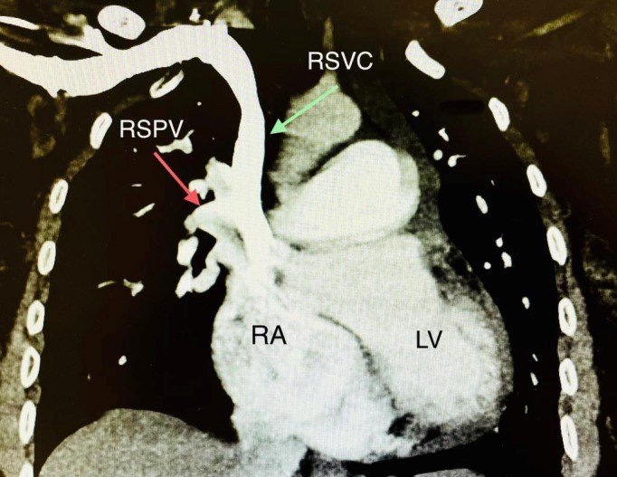
Detecting dilated coronary sinus when assessing patients in an acute emergency with point-of-care ultrasound (POCUS) is important for differential diagnosis, including the detection of persistent left superior vena cava (PLSVC) and right ventricular dysfunction. Cardiac POCUS with agitated saline injections through the left and right antecubital veins is a simple bedside test to make the diagnosis. We present a 42-year-old woman with first-time rapid atrial flutter in whom POCUS confirmed the presence of dilated coronary sinus and PLSVC.
Keywords: arrhythmia; echocardiography.
Figures
Similar articles
Rio PP, Mumpuni H, Anggrahini DW, Dinarti LK.Clin Case Rep. 2017 Mar 17;5(5):587-590. doi: 10.1002/ccr3.883. eCollection 2017 May.PMID: 28469854 Free PMC article.
Mousa TM, Akinseye OA, Kerwin TC, Akinboboye OO.Am J Case Rep. 2015 Aug 11;16:528-31. doi: 10.12659/AJCR.894394.PMID: 26262994 Free PMC article.
Sheikh AS, Mazhar S.Echocardiography. 2014 May;31(5):674-9. doi: 10.1111/echo.12514. Epub 2014 Jan 24.PMID: 24460570 Review.
Kolski BC, Khadivi B, Anawati M, Daniels LB, Demaria AN, Blanchard DG.Echocardiography. 2011 Sep;28(8):829-32. doi: 10.1111/j.1540-8175.2011.01445.x. Epub 2011 Aug 9.PMID: 21827538
Gonzalez-Juanatey C, Testa A, Vidan J, Izquierdo R, Garcia-Castelo A, Daniel C, Armesto V.Clin Cardiol. 2004 Sep;27(9):515-8. doi: 10.1002/clc.4960270909.PMID: 15471164 Free PMC article. Review.
KMEL References
References
-
- Labovitz A J, Noble V E, Bierig M, Goldstein S A, Jones R, Kort S, Porter T R, Spencer K T, Tayal V S, Wei K. Focused cardiac ultrasound in the emergent setting: a consensus statement of the American Society of Echocardiography and American College of Emergency Physicians. J AmSoc Echocardiogr. 2010;23(12):1225–1230. - PubMed
-
- Berger F, Vogel M, Kramer A, V Alexi-Meskishvili, Weng Y, Lange P E, Hetzer R. Incidence of atrial flutter/fibrillation in adults with atrial septal defect before and after surgery. Ann Thorac Surg. 1999;68:75–75. - PubMed
-
- Jost C H Attenhofer, Connolly H M, Danielson G K, Bailey K R, Schaff H V, Shen W K, Warnes C A, Seward J B, Puga F J, Tajik A J. Sinus venosus atrial septal defect: long-term postoperative outcome for 115 patients. Circulation. 2005;112:1953. - PubMed
-
- Wenger N K, Lloyd-Jones D M, Elkind Msv, Fonarow G C, Warner J J, Alger H M, Cheng S, Kinzy C, Hall J L, Roger V L, Association American Heart. Circulation. 23. Vol. 145. American Heart Association; 2022. Call to Action for Cardiovascular Disease in Women: Epidemiology, Awareness, Access, and Delivery of Equitable Health Care: A Presidential Advisory from the American Heart Association; pp. e1059–e1071. - DOI - PubMed


