Point-of-Care Ultrasound Can Suggest COVID-19
Affiliations
Affiliations
- Internal Medicine Department, Ahmadi Hospital - Kuwait Oil Company, Kuwait.
Abstract
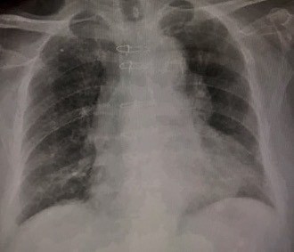
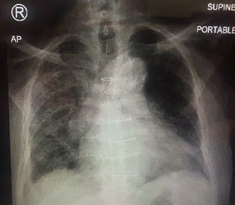
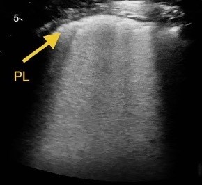
Coronavirus disease 2019 (COVID-19) is caused by severe acute respiratory syndrome coronavirus 2 (SARS-CoV-2) and the World Health Organization (WHO) declared it a pandemic on 11 March 2020. Point-of-care ultrasound (POCUS) is a real-time bedside tool used by physicians to guide rapid, focused and accurate evaluation in order to identify or rule out various pathologies. We describe the case of an elderly man who had fallen at home 3 days previously and was hypoxic at presentation to the emergency department (ED). POCUS in the ED helped to identify a combination of lung and vascular involvement that indicated COVID-19 infection, which was confirmed by a laboratory test.
Learning points: COVID-19 is a contagious disease caused by SARS-CoV-2 that attacks endothelial cells and most organs, resulting in different manifestations and clinical scenarios.Point-of-care ultrasound in the emergency room including lung ultrasound (LUS) and focused echocardiography (FECHO) can be useful in identifying pulmonary and vascular manifestations of COVID-19 disease during the current pandemic.Characteristic LUS signs suggesting bilateral interstitial pneumonia in addition to signs of acute right ventricular strain suggesting pulmonary embolism on FECHO raised the suspicion of COVID-19 infection in our patient.
Keywords: B lines; COVID-19; POCUS; flashing phenomenon; pulmonary embolism.
Conflict of interest statement
Conflicts of Interests: The Authors declare that there are no competing interests.
Figures
Similar articles
Lung Ultrasound Findings in COVID-19: A Descriptive Retrospective Study.
Omer T, Cousins C, Lynch T, Le NN, Sajed D, Mailhot T.Cureus. 2022 Mar 21;14(3):e23375. doi: 10.7759/cureus.23375. eCollection 2022 Mar.PMID: 35475095 Free PMC article.
Bianchi S, Savinelli C, Paolucci E, Pelagatti L, Sibona E, Fersini N, Buggea M, Tozzi C, Allescia G, Paolini D, Lanigra M.Intern Emerg Med. 2022 Jan;17(1):193-204. doi: 10.1007/s11739-021-02643-w. Epub 2021 Apr 21.PMID: 33881727 Free PMC article.
Lung point-of-care (POCUS) ultrasound in a pediatric COVID-19 case.
Alilio PM, Ebeling-Koning NE, Roth KR, Desai T.Radiol Case Rep. 2020 Nov;15(11):2314-2318. doi: 10.1016/j.radcr.2020.09.007. Epub 2020 Sep 7.PMID: 32922585 Free PMC article.
Istvan-Adorjan S, Ágoston G, Varga A, Cotoi OS, Frigy A.Anatol J Cardiol. 2020 Aug;24(2):76-80. doi: 10.14744/AnatolJCardiol.2020.33645.PMID: 32749247 Free PMC article. Review.
Are Lung Ultrasound Findings in COVID-19 Pneumonia Typical or Specific?
Volpicelli G, Cardinale L, Fraccalini T.Praxis (Bern 1994). 2021 Jun;110(8):421-425. doi: 10.1024/1661-8157/a003696.PMID: 34107756 Review.
Cited by
Zaalouk TM, Bitar ZI, Maadarani OS, Ragab Elshabasy RD.Clin Case Rep. 2021 Mar 28;9(5):e04075. doi: 10.1002/ccr3.4075. eCollection 2021 May.PMID: 34084496 Free PMC article.


