Normal chest X-ray should not mislead
Affiliations
Affiliations
- Department of Internal Medicine, KOC Hospital, Fahahil, Kuwait.
Abstract
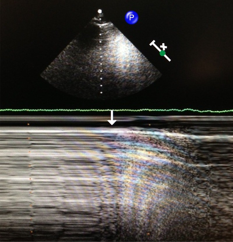
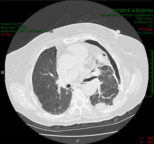
A lung ultrasound (US) can be routinely performed at the bedside by a trained intensive care unit physician and may provide accurate information about a lung's status that has diagnostic and therapeutic relevance. Oesophageal perforations are rare, and due to the rarity of this type of perforation and its non-specific presentation, the diagnosis and treatment are delayed, leading to a high mortality rate. We present a 70-year-old woman with a postoesophagoscopy perforated oesophagus. Lung US detected pneumothorax and mild pleural effusion that were not present on the postoperative chest X-ray. The early detection of the perforation led to a good outcome.
Figures
Similar articles
Nouel O, Rouanet JP, Lichtenstein H, Bloch P, Cugnenc PH, Monnier JP.J Radiol Electrol Med Nucl. 1978 Feb;59(2):113-8.PMID: 641880 French.
Instrumental perforations of the oesophagus.
Moghissi K.Br J Hosp Med. 1988 Mar;39(3):231-6.PMID: 3359096
[Therapeutic case of left pyopneumothorax following esophageal perforation caused by esophagoscopy].
Nakamura Y.Geka Chiryo. 1966 Dec;15(6):751-5.PMID: 6013679 Japanese. No abstract available.
Clinical review: Bedside lung ultrasound in critical care practice.
Bouhemad B, Zhang M, Lu Q, Rouby JJ.Crit Care. 2007;11(1):205. doi: 10.1186/cc5668.PMID: 17316468 Free PMC article. Review.
Phillips LG Jr, Cunningham J.Radiol Clin North Am. 1984 Sep;22(3):607-13.PMID: 6382422 Review.
References
https://pubmed.ncbi.nlm.nih.gov/

