Systematic heterogeneity of fractional vesicle pool sizes and release rates of hippocampal synapses
Affiliations
Affiliations
- Department of Psychiatry and Psychotherapy, University of Erlangen-Nuremberg, Erlangen, Germany.
- Department of Physiology, Faculty of Medicine, Jabriya, Kuwait University, Safat, Kuwait.
- European Neuroscience Institute Göttingen, Deutsche Forschungsgemeinschaft Research Center for Molecular Physiology of the Brain/Excellence Cluster 171, Göttingen, Germany.
- Department of Psychiatry and Psychotherapy, University of Erlangen-Nuremberg, Erlangen, Germany. Electronic address: teja.groemer@uk-erlangen.de.
Abstract
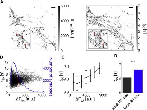
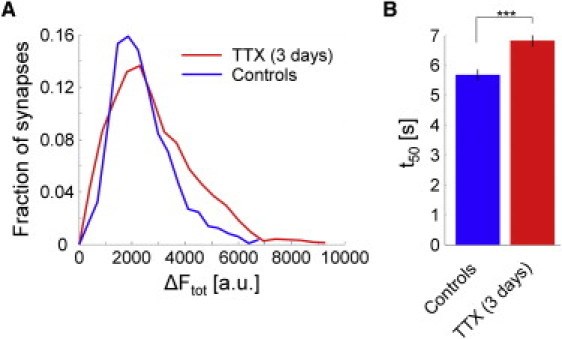
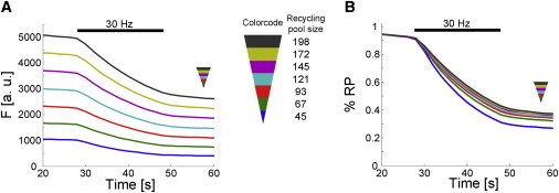
Hippocampal neurons in tissue culture develop functional synapses that exhibit considerable variation in synaptic vesicle content (20-350 vesicles). We examined absolute and fractional parameters of synaptic vesicle exocytosis of individual synapses. Their correlation to vesicle content was determined by activity-dependent discharge of FM-styryl dyes. At high frequency stimulation (30 Hz), synapses with large recycling pools released higher amounts of dye, but showed a lower fractional release compared to synapses that contained fewer vesicles. This effect gradually vanished at lower frequencies when stimulation was triggered at 20 Hz and 10 Hz, respectively. Live-cell antibody staining with anti-synaptotagmin-1-cypHer 5, and overexpression of synaptopHluorin as well as photoconversion of FM 1-43 followed by electron microscopy, consolidated the findings obtained with FM-styryl dyes. We found that the readily releasable pool grew with a power function with a coefficient of 2/3, possibly indicating a synaptic volume/surface dependency. This observation could be explained by assigning the rate-limiting factor for vesicle exocytosis at high frequency stimulation to the available active zone surface that is proportionally smaller in synapses with larger volumes.
Figures
Similar articles
Functional Nanoscale Imaging of Synaptic Vesicle Cycling with Superfast Fixation.
Schikorski T.Methods Mol Biol. 2016;1474:343-58. doi: 10.1007/978-1-4939-6352-2_22.PMID: 27515092
Mechanisms of synaptic vesicle recycling illuminated by fluorescent dyes.
Cousin MA, Robinson PJ.J Neurochem. 1999 Dec;73(6):2227-39. doi: 10.1046/j.1471-4159.1999.0732227.x.PMID: 10582580 Review.
Development of vesicle pools during maturation of hippocampal synapses.
Mozhayeva MG, Sara Y, Liu X, Kavalali ET.J Neurosci. 2002 Feb 1;22(3):654-65. doi: 10.1523/JNEUROSCI.22-03-00654.2002.PMID: 11826095 Free PMC article.
Welzel O, Tischbirek CH, Kornhuber J, Groemer TW.J Neurosci Methods. 2012 Apr 15;205(2):258-64. doi: 10.1016/j.jneumeth.2012.01.011. Epub 2012 Jan 28.PMID: 22306057
Imaging synaptic vesicle recycling by staining and destaining vesicles with FM dyes.
Hoopmann P, Rizzoli SO, Betz WJ.Cold Spring Harb Protoc. 2012 Jan 1;2012(1):77-83. doi: 10.1101/pdb.prot067603.PMID: 22194270 Review.
Cited by
Guhathakurta D, Akdaş EY, Fejtová A, Weiss EM.Front Bioinform. 2022 Feb 15;2:814081. doi: 10.3389/fbinf.2022.814081. eCollection 2022.PMID: 36304276 Free PMC article.
Biasetti L, Rey S, Fowler M, Ratnayaka A, Fennell K, Smith C, Marshall K, Hall C, Vargas-Caballero M, Serpell L, Staras K.Cereb Cortex. 2023 Feb 7;33(4):1263-1276. doi: 10.1093/cercor/bhac134.PMID: 35368053 Free PMC article.
Nanoscale Remodeling of Functional Synaptic Vesicle Pools in Hebbian Plasticity.
Rey S, Marra V, Smith C, Staras K.Cell Rep. 2020 Feb 11;30(6):2006-2017.e3. doi: 10.1016/j.celrep.2020.01.051.PMID: 32049027 Free PMC article.
Henkel AW, Mouihate A, Welzel O.Front Neurosci. 2019 Oct 2;13:1047. doi: 10.3389/fnins.2019.01047. eCollection 2019.PMID: 31632237 Free PMC article.
Chen S, Yu C, Rong L, Li CH, Qin X, Ryu H, Park H.Front Mol Neurosci. 2018 Dec 24;11:478. doi: 10.3389/fnmol.2018.00478. eCollection 2018.PMID: 30618623 Free PMC article.


