The suitability of panoramic radiographs for clinical decision making regarding root angulation compared to cone-beam computed tomography
Affiliations
Affiliations
- Department of Developmental and Preventive Sciences, Faculty of Dentistry, Kuwait University, P.O. Box 24923, 13110, Safat, Kuwait. athbi.ortho@icloud.com.
- Department of Developmental and Preventive Sciences, Faculty of Dentistry, Kuwait University, P.O. Box 24923, 13110, Safat, Kuwait.
- Department of Diagnostic Sciences, Faculty of Dentistry, Kuwait University, Safat, Kuwait.
- Department of Orthodontics, Brodie Craniofacial Endowed Chair, College of Dentistry, University of Illinois at Chicago, Chicago, USA.
Abstract
Background: The study compared clinical decisions regarding root angulation correction and root proximity based on the interpretation of Panoramic (PAN) versus Cone-Beam Computed Tomography (CBCT) images.
Methods: A total of 864 teeth from 36 existing, radiographic patient records at a university dental clinic with concurrent PAN and CBCT images were assessed using PANs, then using CBCTs in a blinded manner by two orthodontists. Teeth were rated regarding the need for root repositioning, the direction of repositioning and existence of root proximity. Frequencies, rating time and intra- and inter-examiner Cohen's Kappa were calculated.
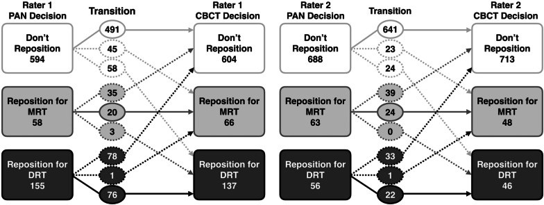
Results: There was 73.7-84.5% agreement between PAN-based and CBCT-based orthodontists' decisions regarding the need to reposition roots. Root proximity was more frequently reported on PANs than CBCTs by one examiner (p = 0.001 and p = 0.168). Both PANs and CBCTs had moderate to substantial intra-examiner, within-radiograph-type reliability with Kappa values of 0.686-0.79 for PANs, and 0.661 for CBCTs (p < 0.001). Inter-examiner and inter-radiograph-type Kappa values ranged from 0.414 to 0.51 (p < 0.001). Using CBCT decisions as a reference, 78.9% of PAN decisions were coincident, 9.3% would have been repositioned on CBCT but not on PAN, 11.3% would not have been repositioned on CBCT but were on PAN, and 0.3% would have been repositioned in the opposite direction on CBCT versus PAN. Additionally, CBCT images required more time per tooth to assess than PANs (p < 0.001).
Conclusions: PAN-based clinical decisions regarding root angulation had comparable statistical reliability and substantial agreement with CBCT-based clinical decisions.
Keywords: Bracket repositioning; Cone-beam CT; Panoramic radiograph; Root angulation; Root proximity.
Conflict of interest statement
None of the authors has any conflict of interest to disclose related to this work.
Figures
Similar articles
Alqerban A, Jacobs R, Fieuws S, Willems G.Eur J Orthod. 2011 Feb;33(1):93-102. doi: 10.1093/ejo/cjq034.PMID: 21270321
Bouwens DG, Cevidanes L, Ludlow JB, Phillips C.Am J Orthod Dentofacial Orthop. 2011 Jan;139(1):126-32. doi: 10.1016/j.ajodo.2010.05.016.PMID: 21195286 Free PMC article.
Van Elslande D, Heo G, Flores-Mir C, Carey J, Major PW.Am J Orthod Dentofacial Orthop. 2010 Apr;137(4 Suppl):S94-9. doi: 10.1016/j.ajodo.2009.02.028.PMID: 20381767
Barakaat AA, Maaz M, Sukhia RH, Fida M.Int Orthod. 2023 May 8;21(3):100757. doi: 10.1016/j.ortho.2023.100757. Online ahead of print.PMID: 37163932
Unexpected artefacts and occult pathologies under CBCT.
Lombardo L, Arreghini A, Guarneri MP, Lauritano D, Nardone M, Siciliani G.Oral Implantol (Rome). 2017 Sep 27;10(2):97-104. doi: 10.11138/orl/2017.10.2.097. eCollection 2017 Apr-Jun.PMID: 29876034 Free PMC article. Review.
KMEL References
References
-
- Orthodontics TABo. Grading System for Dental Casts and Panoramic Radiographs. 2012; https://www.americanboardortho.com/media/1191/grading-system-casts-radio....
-
- Bujang MA, Baharum N. Guidelines of the minimum sample size requirements for Kappa agreement test. Epidemiol Biostat Public Health. 2017;14(2):e12267.

