Root and Canal Morphology of Mandibular Premolar Teeth in a Kuwaiti Subpopulation: A CBCT Clinical Study
Affiliations
Affiliations
- Department of Farwania Dental, Ministry of Health, Kuwait City, Kuwait.
- Department of Restorative Dentistry, Riyadh Elm University, College of Dentistry, Riyadh, Saudi Arabia.
- Department of Preventive Dental Sciences Biostatistics, King Saud University, College of Dentistry, Riyadh, Saudi Arabia.
- Department of Oral and Maxillofacial Surgery Diagnostic Sciences, Riyadh Elm University, College of Dentistry, Riyadh, Saudi Arabia.
Abstract
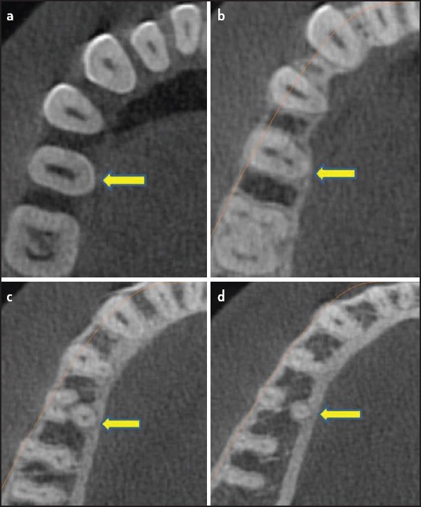
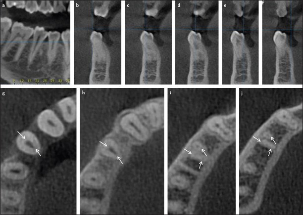
Objective: To study the root and root canal morphology of mandibular premolars in a Kuwaiti subpopulation using cone-beam computed tomography (CBCT).
Methods: 152 CBCT images were obtained from the radiology department archives of four dental centers in Kuwait. A total of 476 mandibular premolar teeth were analyzed by two observers. The number of roots, root canal configuration types and canal curvature measurements were examined. The relationship between sex, tooth position, and incidence of an additional canal were compared using the chi-square test, and the level of significance was set at 0.05 (P=0.05).
Results: The number of roots in mandibular first premolars was one in 73.9%, two in 24.9%, three and four in 1.2%. On the other hand, the number of roots in mandibular second premolars was one in 79.2% and two in 20.8%. Based on Vertucci's classification system, 18.7% of the teeth were type II followed by type VI (14.3%). The majority of the examined teeth were straight (74.8%) and the incidence of distal root angulation was about 21%. Canal configurations not included in the Vertucci classification were reported in 102 teeth (21.4%). Variability was significantly higher in the second premolars compared to first premolar (P<0.05).
Conclusion: The Kuwaiti population has complex root canal morphology in mandibular premolar teeth.
Conflict of interest statement
Conflict of Interest: No conflict of interest.
Figures
Similar articles
Karobari MI, Iqbal A, Syed J, Batul R, Adil AH, Khawaji SA, Howait M, Khattak O, Noorani TY.BMC Oral Health. 2023 May 15;23(1):291. doi: 10.1186/s12903-023-03002-1.PMID: 37189077 Free PMC article.
Ok E, Altunsoy M, Nur BG, Aglarci OS, Çolak M, Güngör E.Acta Odontol Scand. 2014 Nov;72(8):701-6. doi: 10.3109/00016357.2014.898091. Epub 2014 May 15.PMID: 24832561
Saber SEDM, Ahmed MHM, Obeid M, Ahmed HMA.Int Endod J. 2019 Mar;52(3):267-278. doi: 10.1111/iej.13016. Epub 2018 Nov 8.PMID: 30225932
Aksoy U, Aksoy S, Orhan K.Microsc Res Tech. 2018 Mar;81(3):308-314. doi: 10.1002/jemt.22980. Epub 2017 Dec 29.PMID: 29285826 Review.
The root and root canal morphology of the human mandibular second premolar: a literature review.
Cleghorn BM, Christie WH, Dong CC.J Endod. 2007 Sep;33(9):1031-7. doi: 10.1016/j.joen.2007.03.020. Epub 2007 Jun 5.PMID: 17931927 Review.
Cited by
Alazemi HS, Al-Nazhan SA, Aldosimani MA.Saudi Dent J. 2023 May;35(4):345-353. doi: 10.1016/j.sdentj.2023.03.008. Epub 2023 Mar 24.PMID: 37251720 Free PMC article.
Al-Rammahi HM, Chai WL, Nabhan MS, Ahmed HMA.BMC Oral Health. 2023 May 29;23(1):339. doi: 10.1186/s12903-023-03036-5.PMID: 37248469 Free PMC article.
Karobari MI, Iqbal A, Syed J, Batul R, Adil AH, Khawaji SA, Howait M, Khattak O, Noorani TY.BMC Oral Health. 2023 May 15;23(1):291. doi: 10.1186/s12903-023-03002-1.PMID: 37189077 Free PMC article.
KMEL References
References
-
- Vertucci FJ. Root canal morphology of mandibular premolars. J Am Dent Assoc. 1978;97(1):47–50. - PubMed
-
- Vertucci FJ. Root canal anatomy of the human permanent teeth. Oral Surg Oral Med Oral Pathol. 1984;58(5):589–99. - PubMed
-
- Cantatore G, Berutti E, Castellucci A. Missed anatomy:Frequency and clinical impact. Endod Topics. 2006;15(1):3–31.
-
- Slowey RR. Root canal anatomy. Road map to successful endodontics. Dent Clin North Am. 1979;23(4):555–573. - PubMed
-
- Lee YY, Yeh PY, Pai SF, Yang SF. Maxillary first molar with six canals. J Dent Sci. 2009;4(4):198–201.
-
- Jha P, Nikhil V, Arora V, Jha M. The root and root canal morphology of the human mandibular premolars:A literature review. J Rest Dent. 2013;1(1):3–10.
-
- Cleghorn BM, Christie WH, Dong CC. The root and root canal morphology of the human mandibular first premolar:a literature review. J Endod. 2007;33(5):509–16. - PubMed
-
- Al-Fouzan KS. The microscopic diagnosis and treatment of a mandibular second premolar with four canals. Int Endod J. 2001;34(5):406–10. - PubMed
-
- Al-Abdulwahhab B, Al-Nazhan S. Root canal treatment of mandibular second premolar with four root canals. Saudi Endod J. 2015;5(3):196–8.
-
- Fan B, Yang J, Gutmann JL, Fan M. Root canal systems in mandibular first premolars with C-shaped root configurations. Part I:Microcomputed tomography mapping of the radicular groove and associated root canal cross-sections. J Endod. 2008;34(11):1337–41. - PubMed
-
- Lu TY, Yang SF, Pai SF. Complicated root canal morphology of mandibular first premolar in a Chinese population using the cross section method. J Endod. 2006;32(10):932–6. - PubMed
-
- Pineda F, Kuttler Y. Mesiodistal and buccolingual roentgenographic investigation of 7,275 root canals. Oral Surg Oral Med Oral Pathol. 1972;33(1):101–10. - PubMed
-
- Gulabivala K, Aung TH, Alavi A, Ng YL. Root and canal morphology of Burmese mandibular molars. Int Endod J. 2001;34(5):359–70. - PubMed
-
- Sert S, Bayirli GS. Evaluation of the root canal configurations of the mandibular and maxillary permanent teeth by gender in the Turkish population. J Endod. 2004;30(6):391–8. - PubMed
-
- Kim E, Fallahrastegar A, Hur YY, Jung IY, Kim S, Lee SJ. Difference in root canal length between Asians and Caucasians. Int Endod J. 2005;38(3):149–51. - PubMed
-
- Awawdeh L, Abdullah H, Al-Qudah A. Root form and canal morphology of Jordanian maxillary first premolars. J Endod. 2008;34(8):956–61. - PubMed
-
- Zaatar EI, al-Kandari AM, Alhomaidah S, al-Yasin IM. Frequency of endodontic treatment in Kuwait:radiographic evaluation of 846 endodontically treated teeth. J Endod. 1997;23(7):453–6. - PubMed
-
- Zaatar EI, al Anizi SA, al Duwairi Y. A study of the dental pulp cavity of mandibular first permanent molars in the Kuwaiti population. J Endod. 1998;24(2):125–7. - PubMed
-
- Pattanshetti N, Gaidhane M, Al Kandari AM. Root and canal morphology of the mesiobuccal and distal roots of permanent first molars in a Kuwait population-a clinical study. Int Endod J. 2008;41(9):755–62. - PubMed
-
- Chourasia HR, Boreak N, Tarrosh MY, Mashyakhy M. Root canal morphology of mandibular first premolars in Saudi Arabian southern region subpopulation. Saudi Endod J. 2017;7(2):77–81.
-
- Boschetti E, Silva-Sousa YTC, Mazzi-Chaves JF, Leoni GB, Versiani MA, Pécora JD, et al. Micro-CT Evaluation of Root and Canal Morphology of Mandibular First Premolars with Radicular Grooves. Braz Dent J. 2017;28(5):597–603. - PubMed
-
- Ok E, Altunsoy M, Nur BG, Aglarci OS, Çolak M, Güngör E. A cone-beam computed tomography study of root canal morphology of maxillary and mandibular premolars in a Turkish population. Acta Odontol Scand. 2014;72(8):701–6. - PubMed
-
- Hashimoto K, Kawashima S, Kameoka S, Akiyama Y, Honjoya T, Ejima K, et al. Comparison of image validity between cone beam computed tomography for dental use and multidetector row helical computed tomography. Dentomaxillofac Radiol. 2007;36(8):465–71. - PubMed
-
- Schneider SW. A comparison of canal preparations in straight and curved root canals. Oral Surg Oral Med Oral Pathol. 1971;32(2):271–5. - PubMed
-
- Nelson SJ. Wheeler's dental anatomy physiology and occlusion. 10th ed. Elsevier Health Sciences; 2014.
-
- Peiris R. Root and canal morphology of human permanent teeth in a Sri Lankan and Japanese population. Anthropological Science. 2008;116(2):123–33.
-
- Hertzog MA. Considerations in determining sample size for pilot studies. Res Nurs Health. 2008;31(2):180–91. - PubMed
-
- Isaac S, Michael WB. Handbook in research and evaluation. San Diego, CA: Educational and Industrial Testing Services; 1995. p. 101.
-
- Yoshioka T, Villegas JC, Kobayashi C, Suda H. Radiographic evaluation of root canal multiplicity in mandibular first premolars. J Endod. 2004;30(2):73–4. - PubMed
-
- Omer OE, Al Shalabi RM, Jennings M, Glennon J, Claffey NM. A comparison between clearing and radiographic techniques in the study of the root-canal anatomy of maxillary first and second molars. Int Endod J. 2004;37(5):291–6. - PubMed
-
- Neelakantan P, Subbarao C, Subbarao CV. Comparative evaluation of modified canal staining and clearing technique cone-beam computed tomography peripheral quantitative computed tomography spiral computed tomography and plain and contrast medium-enhanced digital radiography in studying root canal morphology. J Endod. 2010;36(9):1547–51. - PubMed
-
- Liao Q, Han JL, Xu X. Analysis of canal morphology of mandibular first premolar [Article in Chinese] Shanghai Kou Qiang Yi Xue. 2011;20(5):517–21. - PubMed
-
- Sousa TO, Haiter-Neto F, Nascimento EHL, Peroni LV, Freitas DQ, Hassan B. Diagnostic Accuracy of Periapical Radiography and Cone-beam Computed Tomography in Identifying Root Canal Configuration of Human Premolars. J Endod. 2017;43(7):1176–9. - PubMed
-
- Patel S, Durack C, Abella F, Shemesh H, Roig M, Lemberg K. Cone beam computed tomography in Endodontics - a review. Int Endod J. 2015;48(1):3–15. - PubMed
-
- Bürklein S, Heck R, Schäfer E. Evaluation of the Root Canal Anatomy of Maxillary and Mandibular Premolars in a Selected German Population Using Cone-beam Computed Tomographic Data. J Endod. 2017;43(9):1448–52. - PubMed
-
- Martins JNR, Francisco H, Ordinola-Zapata R. Prevalence of C-shaped Configurations in the Mandibular First and Second Premolars:A Cone-beam Computed Tomographic In Vivo Study. J Endod. 2017;43(6):890–5. - PubMed
-
- Vertucci FJ. Root canal morphology and its relationship to endodontic procedures. Endod Topics. 2005;10(1):3–29.
-
- Kartal N, Yanikoğlu FC. Root canal morphology of mandibular incisors. J Endod. 1992;18(11):562–4. - PubMed
-
- Peiris HR, Pitakotuwage TN, Takahashi M, Sasaki K, Kanazawa E. Root canal morphology of mandibular permanent molars at different ages. Int Endod J. 2008;41(10):828–35. - PubMed
-
- Al-Qudah AA, Awawdeh LA. Root and canal morphology of mandibular first and second molar teeth in a Jordanian population. Int Endod J. 2009;42(9):775–84. - PubMed
-
- Ahmed HMA, Versiani MA, De-Deus G, Dummer PMH. A new system for classifying root and root canal morphology. Int Endod J. 2017;50(8):761–70. - PubMed
-
- Gu YC, Zhang YP, Liao ZG, Fei XD. A micro-computed tomographic analysis of wall thickness of C-shaped canals in mandibular first premolars. J Endod. 2013;39(8):973–6. - PubMed
-
- Reddy SJ, Chandra PV, Santoshi L, Reddy GV. Endodontic management of two-rooted mandibular premolars using spiral computed tomography:a report of two cases. J Contemp Dent Pract. 2012;13(6):908–13. - PubMed
-
- Fan B, Cheung GS, Fan M, Gutmann JL, Bian Z. C-shaped canal system in mandibular second molars:Part I--Anatomical features. J Endod. 2004;30(12):899–903. - PubMed
-
- Kotoku K. Morphological studies on the roots of Japanese mandibular second molars [Article in Japanese] Shikwa Gakuho. 1985;85(1):43–64. - PubMed
-
- Woelfel J, Scheid R. Dental anatomy:its relevance to dentistry. 6th ed. Philadelphia: Lippincott Williams &Wilkins; 2002.
-
- Lee KW, Lee EC, Poon KY. Palato-gingival grooves in maxillary incisors. A possible predisposing factor to localised periodontal disease. Br Dent J. 1968;124(1):14–8. - PubMed
-
- Wasserstein A, Brezniak N, Shalish M, Heller M, Rakocz M. Angular changes and their rates in concurrence to developmental stages of the mandibular second premolar. Angle Orthod. 2004;74(3):332–6. - PubMed
-
- Shalish M, Peck S, Wasserstein A, Peck L. Malposition of unerupted mandibular second premolar associated with agenesis of its antimere. Am J Orthod Dentofacial Orthop. 2002;121(1):53–6. - PubMed
-
- Van der Linden FPGM, Duterloo HS. Development of the Human Dentition:An Atlas. Hagerstown: Harper &Row; 1976. pp. 204–5.
-
- Mckee IW, Glover KE, Williamson PC, Lam EW, Heo G, Major PW. The effect of vertical and horizontal head positioning in panoramic radiography on mesiodistal tooth angulations. Angle Orthod. 2001;71(6):442–51. - PubMed
-
- Cleghorn BM, Christie WH, Dong CC. The root and root canal morphology of the human mandibular second premolar:a literature review. J Endod. 2007;33(9):1031–7. - PubMed
-
- Khedmat S, Assadian H, Saravani AA. Root canal morphology of the mandibular first premolars in an Iranian population using cross-sections and radiography. J Endod. 2010;36(2):214–7. - PubMed
-
- Lombart B, Michonneau JC. -Premolar anatomy and endodontic treatment. Rev Belge Med Dent. 2005;60(4):322–36. - PubMed


