A rabbit model for experimental alveolar cleft grafting
Affiliations
Affiliations
- Department of Cranio-Maxillofacial Surgery, Maastricht University Medical Center, P. Debyelaan, Postbus 5800, 6202 AZ, Maastricht, The Netherlands. mkamal@ukaachen.de.
- Department of Oral and Maxillofacial Surgery, RWTH Aachen University, Pauwelsstraße 30, 52074, Aachen, Germany. mkamal@ukaachen.de.
- Department of Surgical Sciences, Faculty of Dentistry, Health Sciences Center, Kuwait University, 13110, Safat, Kuwait.
- Institute for Laboratory Animal Science and Experimental Surgery, RWTH Aachen University, Pauwelsstraße 30, 52074, Aachen, Germany.
- Department of Oral and Maxillofacial Surgery, RWTH Aachen University, Pauwelsstraße 30, 52074, Aachen, Germany.
- Department of Experimental Molecular Imaging, RWTH Aachen University, Pauwelsstraße 30, 52074, Aachen, Germany.
- Department of Cranio-Maxillofacial Surgery, Maastricht University Medical Center, P. Debyelaan, Postbus 5800, 6202 AZ, Maastricht, The Netherlands.
Abstract
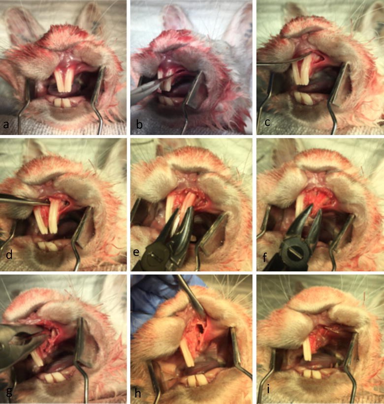
Objectives: The purpose of the present study was to develop an animal model for creating alveolar cleft defects with properly simulated clinical defect environment for tissue-engineered bone-substitute materials testing without compromising the health of the animal. Cleft creation surgery was aimed at creating a complete alveolar cleft with a wide bone defect with an epithelial lining (oral mucosa) overlying the cleft defect.
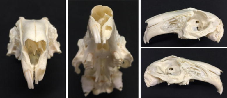
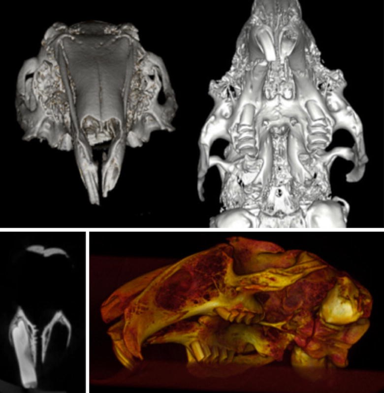
Methods: A postmortem skull of a New Zealand White (NZW) rabbit skull (Oryctolagus cuniculus) underwent an osteological and imaging survey. A pilot postmortem surgery was conducted to confirm the feasability of a surgical procedure and the defect was also radiologically confirmed and illustrated with micro-computed tomography. Then, a surgical in vivo model was tested and evaluated in 16 (n = 16) 8-week-old NZW rabbits to create in vivo alveolar cleft creation surgery.
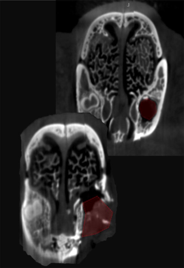
Results: Clinical examination and imaging analysis 8 weeks after cleft creation surgery revealed the establishment of a wide skeletal defect extending to the nasal mucosa simulating alveolar clefts in all of the rabbits.
Conclusions: Our surgical technique was successful in creating a sizable and predictable model for bone grafting material testing. The model allows for simulating the cleft site environment and can be used to evaluate various bone grafting materials in regard to efficacy of osteogenesis and healing potential without compromising the health of the animal.
Keywords: Animal testing; Cleft lip and palate; Grafting; Rabbit; Tissue-engineering.
Figures
Similar articles
Kamal M, Al-Obaidly S, Lethaus B, Bartella AK.Clin Exp Dent Res. 2022 Dec;8(6):1331-1340. doi: 10.1002/cre2.644. Epub 2022 Aug 7.PMID: 35933723 Free PMC article.
Kamal M, Andersson L, Tolba R, Al-Asfour A, Bartella AK, Gremse F, Rosenhain S, Hölzle F, Kessler P, Lethaus B.J Transl Med. 2017 Dec 23;15(1):263. doi: 10.1186/s12967-017-1369-3.PMID: 29274638 Free PMC article.
New technique for creating permanent experimental alveolar clefts in a rabbit model.
el-Bokle D, Smith SJ, Germane N, Sharawy M.Cleft Palate Craniofac J. 1993 Nov;30(6):542-7. doi: 10.1597/1545-1569_1993_030_0542_ntfcpe_2.3.co_2.PMID: 8280731
Osteoplasty of the alveolar cleft defect.
Rychlik D, Wójcicki P, Koźlik M.Adv Clin Exp Med. 2012 Mar-Apr;21(2):255-62.PMID: 23214291 Review.
Secondary bone grafting for alveolar cleft in children with cleft lip or cleft lip and palate.
Guo J, Li C, Zhang Q, Wu G, Deacon SA, Chen J, Hu H, Zou S, Ye Q.Cochrane Database Syst Rev. 2011 Jun 15;(6):CD008050. doi: 10.1002/14651858.CD008050.pub2.PMID: 21678372 Review.
Cited by
Kamal M, Al-Obaidly S, Lethaus B, Bartella AK.Clin Exp Dent Res. 2022 Dec;8(6):1331-1340. doi: 10.1002/cre2.644. Epub 2022 Aug 7.PMID: 35933723 Free PMC article.
Möhlhenrich SC, Kniha K, Magnuska Z, Chhatwani S, Hermanns-Sachweh B, Gremse F, Hölzle F, Danesh G, Modabber A.Clin Oral Investig. 2022 Sep;26(9):5809-5821. doi: 10.1007/s00784-022-04537-3. Epub 2022 May 14.PMID: 35567639 Free PMC article.
Lee SW, Kim JY, Hong KY, Choi TH, Kim BJ, Kim S.Arch Craniofac Surg. 2021 Oct;22(5):239-246. doi: 10.7181/acfs.2021.00325. Epub 2021 Oct 20.PMID: 34732035 Free PMC article.
Möhlhenrich SC, Kniha K, Magnuska Z, Hermanns-Sachweh B, Gremse F, Hölzle F, Danesh G, Modabber A.Sci Rep. 2021 Jun 30;11(1):13586. doi: 10.1038/s41598-021-93033-x.PMID: 34193933 Free PMC article.
AlOtaibi NM, Dunne M, Ayoub AF, Naudi KB.J Transl Med. 2021 Jun 28;19(1):276. doi: 10.1186/s12967-021-02944-w.PMID: 34183031 Free PMC article.
KMEL References
References
-
- Rychlik D, Wójcicki P, Koźlik M. Osteoplasty of the alveolar cleft defect. Adv Clin Exp Med. 2012;21:255–262. - PubMed
-
- Kawata T, Kohno S, Fujita T, Sugiyama H, Tokimasa C, Kaku M, Tanne K. New biomaterials and methods for craniofacial bone defect: chondroid bone grafts in maxillary alveolar clefts. J Craniofac Genet Dev Biol. 2000;20:49–52. - PubMed
-
- Papadopoulos MA, Papadopulos NA, Jannowitz C, Boettcher P, Henke J, Stolla R, Zeilhofer HF, Kovacs L, Biemer E. Three-dimensional cephalometric evaluation of maxillary growth following in utero repair of cleft lip and alveolar-like defects in the mid-gestational sheep model. Fetal Diagn Ther. 2006;21:105–114. doi: 10.1159/000089059. - DOI - PubMed
-
- Pilanci O, Cinar C, Kuvat SV, Altintas M, Guzel Z, Kilic A. Effects of hydroxyapatite on bone graft resorption in an experimental model of maxillary alveolar arch defects. Arch Clin Exp Surg. 2013;2:170–175. doi: 10.5455/aces.20121018123137. - DOI
-
- Raposo-Amaral CE, Kobayashi GS, Almeida AB, Bueno DF, Freitas FR, Vulcano LC, Passos-Bueno MR, Alonso N. Alveolar osseous defect in rat for cell therapy: preliminary report. Acta Cir Bras. 2010;25:313–317. - PubMed
-
- Sawada Y, Hokugo A, Nishiura A, Hokugo R, Matsumoto N, Morita S, Tabata Y. A trial of alveolar cleft bone regeneration by controlled release of bone morphogenetic protein: an experimental study in rabbits. Oral Surg Oral Med Oral Pathol Oral Radiol Endod. 2009;108:812–820. doi: 10.1016/j.tripleo.2009.06.040. - DOI - PubMed
-
- Wu L-L, Zhao Y, Chen C. Establishment of the animal model with unilateral alveolar cleft and its effect on the nose growth. Zhonghua Zheng Xing Wai Ke Za Zhi. 2010;26:39–42. - PubMed
-
- Igawa HH, Ohura T, Iwao F, Yamamoto Y, Fujioka H. Intrauterine repair of cleft lip in mouse fetuses. Congenit Anom. 1991;31:95–100. doi: 10.1111/j.1741-4520.1991.tb00363.x. - DOI
-
- Wang X, Mabrey JD, Agrawal CM. An interspecies comparison of bone fracture properties. Biomed Mater Eng. 1998;8:1–10. - PubMed
-
- Kolk A, Handschel J, Drescher W, Rothamel D, Kloss F, Blessmann M, Heiland M, Wolff KD, Smeets R. Current trends and future perspectives of bone substitute materials—from space holders to innovative biomaterials. J Craniomaxillofac Surg. 2012;40:706–718. doi: 10.1016/j.jcms.2012.01.002. - DOI - PubMed
-
- Puumanen K, Kellomaki M, Ritsila V, Bohling T, Tormala P, Waris T, Ashammakhi N. A novel bioabsorbable composite membrane of polyactive 70/30 and bioactive glass number 13–93 in repair of experimental maxillary alveolar cleft defects. J Biomed Mater Res B Appl Biomater. 2005;75:25–33. doi: 10.1002/jbm.b.30218. - DOI - PubMed
-
- Directive C. 86/609/EEC of 24 November 1986 on the approximation of laws, regulations and administrative provisions of the Member States regarding the protection of animals used for experimental and other scientific purposes. Off J Eur Commun. 1986;29:L358.


