Prevalence of oral mucosal lesions in patients of the Kuwait University Dental Center
Affiliations
Affiliations
- Department of Diagnostic Sciences, Faculty of Dentistry, Kuwait University, Jabriya, Kuwait.
Abstract
Objectives: The purpose of this study was to determine the number, types, and locations of oral mucosal lesions in patients who attended the Admission Clinic at the Kuwait University Dental Center to determine prevalence and risk factors for oral lesions.
Subjects and methods: Intraoral soft tissue examination was performed on new patients seen between January 2009 and February 2011. The lesions were divided into six major groups: white, red, pigmented, ulcerative, exophytic, and miscellaneous.
Results: Five hundred thirty patients were screened, out of which 308 (58.1%) had one or more lesions. A total of 570 oral lesions and conditions were identified in this study, of which 272 (47.7%) were white, 25 (4.4%) were red, 114 (20.0%) were pigmented, 21 (3.7%) were ulcerative, 108 (18.9%) were exophytic, and 30 (5.3%) were in the miscellaneous group. Overall, Fordyce granules (n = 116; 20.4%) were the most frequently detected condition. A significantly higher (p < 0.001) percentage of older patients (21-40 years and ⩾41 years) had oral mucosal lesions than those in the ⩽20 years age group. A significantly higher (p < 0.01) percentage of smokers had oral mucosal lesions than did nonsmokers. Most of the lesions and conditions were found on the buccal mucosa and gingiva.
Conclusions: White, pigmented, and exophytic lesions were the most common types of oral mucosal lesions found in this study. Although most of these lesions are innocuous, the dentist should be able to recognize and differentiate them from the worrisome lesions, and decide on the appropriate treatment.
Keywords: Kuwait; Oral lesions; Oral mucosa; Prevalence; Screening; Tobacco use.
Figures
Similar articles
Access keysNCBI HomepageMyNCBI HomepageMain ContentMain Navigation
Search:0 results are available, use up and down arrow keys to navigate.Search
SaveEmail
Send to
Display options
Abstract PubMed PMID
full text links
actions
Cite
Collections
share
page navigation
Title & authors Abstract Figures Similar articles Cited by LinkOut - more resources
Saudi Dent J
. 2013 Jul;25(3):111-8.
doi: 10.1016/j.sdentj.2013.05.003. Epub 2013 Jun 13.
Prevalence of oral mucosal lesions in patients of the Kuwait University Dental Center
Mohammad Ali 1, Bobby Joseph, Devipriya Sundaram
Affiliations collapse
Affiliation
- 1Department of Diagnostic Sciences, Faculty of Dentistry, Kuwait University, Jabriya, Kuwait.
- PMID: 24179320
- PMCID: PMC3809497
- DOI: 10.1016/j.sdentj.2013.05.003
Free PMC article
Abstract
Objectives: The purpose of this study was to determine the number, types, and locations of oral mucosal lesions in patients who attended the Admission Clinic at the Kuwait University Dental Center to determine prevalence and risk factors for oral lesions.
Subjects and methods: Intraoral soft tissue examination was performed on new patients seen between January 2009 and February 2011. The lesions were divided into six major groups: white, red, pigmented, ulcerative, exophytic, and miscellaneous.
Results: Five hundred thirty patients were screened, out of which 308 (58.1%) had one or more lesions. A total of 570 oral lesions and conditions were identified in this study, of which 272 (47.7%) were white, 25 (4.4%) were red, 114 (20.0%) were pigmented, 21 (3.7%) were ulcerative, 108 (18.9%) were exophytic, and 30 (5.3%) were in the miscellaneous group. Overall, Fordyce granules (n = 116; 20.4%) were the most frequently detected condition. A significantly higher (p < 0.001) percentage of older patients (21-40 years and ⩾41 years) had oral mucosal lesions than those in the ⩽20 years age group. A significantly higher (p < 0.01) percentage of smokers had oral mucosal lesions than did nonsmokers. Most of the lesions and conditions were found on the buccal mucosa and gingiva.
Conclusions: White, pigmented, and exophytic lesions were the most common types of oral mucosal lesions found in this study. Although most of these lesions are innocuous, the dentist should be able to recognize and differentiate them from the worrisome lesions, and decide on the appropriate treatment.
Keywords: Kuwait; Oral lesions; Oral mucosa; Prevalence; Screening; Tobacco use.
Figures

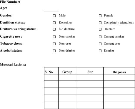
Figure 1
Data collection sheet.

Similar articles
Dental students' ability to detect and diagnose oral mucosal lesions.
Ali MA, Joseph BK, Sundaram DB.J Dent Educ. 2015 Feb;79(2):140-5.PMID: 25640618
El Toum S, Cassia A, Bouchi N, Kassab I.Int J Dent. 2018 May 17;2018:4030134. doi: 10.1155/2018/4030134. eCollection 2018.PMID: 29887889 Free PMC article.
Bhatnagar P, Rai S, Bhatnagar G, Kaur M, Goel S, Prabhat M.J Family Community Med. 2013 Jan;20(1):41-8. doi: 10.4103/2230-8229.108183.PMID: 23723730 Free PMC article.
Prevalence of oral mucosal lesions in alcohol misusers in south London.
Harris CK, Warnakulasuriya KA, Cooper DJ, Peters TJ, Gelbier S.J Oral Pathol Med. 2004 May;33(5):253-9. doi: 10.1111/j.0904-2512.2004.00142.x.PMID: 15078483
Peripheral Exophytic Oral Lesions: A Clinical Decision Tree.
Mortazavi H, Safi Y, Baharvand M, Rahmani S, Jafari S.Int J Dent. 2017;2017:9193831. doi: 10.1155/2017/9193831. Epub 2017 Jul 5.PMID: 28757870 Free PMC article. Review.
Cited by
Epidemiologic and histopathological evaluation of unclassified gingival papules in Urmia, Iran.
Khashabi E, Taram S, Saatloo MV, Farjah GH, Sharifi P, Gobaran ZM.J Oral Maxillofac Pathol. 2023 Jan-Mar;27(1):20-25. doi: 10.4103/jomfp.jomfp_122_21. Epub 2023 Mar 21.PMID: 37234298 Free PMC article.
Mahdani FY, Parmadiati AE, Ernawati DS, Suryanijaya VE, Inastu CR, Radithia D, Ayuningtyas NF, Surboyo MDC, Pratiwi AS, Marsetyo RI.Int Arch Otorhinolaryngol. 2022 Apr 20;26(4):e671-e675. doi: 10.1055/s-0042-1742328. eCollection 2022 Oct.PMID: 36405462 Free PMC article.
Collins JR, Brache M, Ogando G, Veras K, Rivera H.Acta Odontol Latinoam. 2021 Dec 31;34(3):249-256. doi: 10.54589/aol.34/3/249.PMID: 35088812 Free PMC article.
Ogunrinde TJ, Olawale OF.Pan Afr Med J. 2020 Dec 21;37:358. doi: 10.11604/pamj.2020.37.358.22194. eCollection 2020.PMID: 33796172 Free PMC article.
Alsharif MT, Alsharif AT, Krsoum MA, Aljohani MA, Qadiri OM, Alharbi AA, Al-Maweri SA, Warnakulasuriya S, Kassim S.Eur J Dent. 2021 Jul;15(3):509-514. doi: 10.1055/s-0040-1722090. Epub 2021 Feb 23.PMID: 33622006 Free PMC article.


